A new study from the Hebrew University-Hadassah Medical Center has indicated that natural psilocybin extracts may demonstrate superior efficacy to synthetic psilocybin extracts.
Recent years have seen a boom in research into psilocybin for the treatment of mental health conditions such as anxiety and depression.
Many of the clinical trials investigating psilocybin use synthetic extracts rather than natural ones. This is because synthetic extracts will contain psilocybin alone, whereas natural psilocybe mushroom extracts will contain several different compounds such as psilocybin, psilocin, baeocystin and norbaeocystin.
Having multiple compounds can pose a challenge when running clinical trials as identifying which compounds are active and what their impact is becomes difficult to measure, and the concentrations of these compounds can vary depending on factors such as growth conditions and processing techniques.
This makes the standardisation of multi-compound medicines a huge challenge, as medicine consistency, reproducibility and dosing become difficult. However, these are essential factors when it comes to conducting clinical trials and receiving approval for medicines from regulators.
The Entourage Effect
In 2011 Dr Ethan Russo put forward the theory of the Entourage Effect in cannabis.
The cannabis plant contains over 400 different cannabinoids that have so far been identified, such as THC, CBD, CBN and CBG.
Russo hypothesised that these different cannabinoid compounds work synergistically to create a therapeutic effect, as opposed to compounds such as THC or CBD working in isolation.
This hypothesis has been touched on only a few times in the scientific literature in relation to psychedelic mushrooms.
For example, in Dr Jochen Gartz’s 1989 paper ‘Biotransformation of tryptamine derivatives in mycelial cultures of Psilocybe’ which proposed a synergistic relationship between compounds in the mushrooms, and a 2015 paper by Zhuck et al, ‘Research on Acute Toxicity and the Behavioral Effects of Methanolic Extract from Psilocybin Mushrooms and Psilocin in Mice’, which observed that the effect of psychedelic mushroom extracts on mice was much stronger than pure psilocybin.
There has been very limited research on this hypothesis in mushrooms since.
A new study: Natural may outperform synthetic
Now, a research team from Hebrew University-Hadassah Medical Center BrainLabs Center for the Psychedelic Research have compared a natural psilocybin extract to a chemically synthesised version.
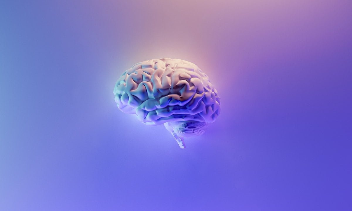
Published in Molecular Psychiatry, results from the study indicate that the natural extract increased the levels of synaptic proteins associated with neuroplasticity in key brain regions, including the frontal cortex, hippocampus, amygdala, and striatum.
The ability of psilocybin to induce neuralplasticity has been indicated as one of the key features that contribute to its therapeutic effects.
The researchers suggest that these new study results indicate that nautral psilocybin extracts may offer unique therapeutic effects that may not be not achievable with synthesised, single-compound psilocybin alone.
Metabolomic analyses also revealed that the natural extract exhibited a distinct metabolic profile associated with oxidative stress and energy production pathways.
The researchers write: “In Western medicine, there has historically been a preference for isolating active compounds rather than utilising extracts, primarily for the sake of gaining better control over dosages and anticipating known effects during treatment. The challenge with working with extracts lay in the inability, in the past, to consistently produce the exact product with a consistent compound profile.
“Contrastingly, ancient medicinal practices, particularly those attributing therapeutic benefits to psychedelic medicine, embraced the use of extracts or entire products, such as consuming the entire mushroom. Although Western medicine has long recognised the “entourage” effect associated with whole extracts, the significance of this approach has gained recent prominence.”
However, compared to cannabis, the researchers suggest that mushroom extracts present a unique case, as they are highly influenced by their growing environment such as substrate, light exposure temperature and more.
“Despite these influences, controlled cultivation allows for the taming of mushrooms, enabling the production of a replicable extract,” the team writes.
The researchers emphasise that this research underscores the superiority of extracts with diverse compounds, and also highlights the feasibility of incorporating them into Western medicine due to the controlled nature of mushroom cultivation.


 Opinion2 years ago
 Insight3 years ago
 Medicinal2 years ago
 Research2 years ago
 Medicinal2 years ago
 Markets & Industry1 year ago
 News3 years ago
 Medicinal2 years ago


